|
|
 |
Parkinson's Disease |
|||
|---|---|---|---|
|
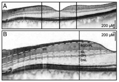
Parkinson Disease (PD) is a progressive neurodegenerative disease. Current research attempts to find protection against progression. Optical coherence tomorgraphy is being used to help develop objective and quantifiable markers of neurodegeneration in PD. The technique of Optical Coherence Tomography is easy, takes only a few minutes, and hence lends itself as a potential biomarker to follow the progression of PD. Potential treatment effects can take advantage of the simplicity of administration and quantification of OCT in PD. Parkinson disease (PD) also affects vision. Several studies using OCT have suggested that the retina (the back of the eye) is affected in PD. The OCT method allows precise quantification of retinal changes in PD. OCT does not pose any discomfort beyond what is experienced by routine eye examinations which involve placing the chin on a stabilizing rest and the pupil is dilated. OCT noninvasively quantifies the thickness of the retinal nerve fiber layer (RNFL). OCT has been studied in several neuro-ophthalmic conditions, including Parkinson’s disease (PD). Recent studies suggest that the quantitative analysis of RNFL can be precisely and noninvasively done by OCT scans and the results suggest that the thickness of RNFL is significantly decreased in patients with PD compared with age-matched controls and the foveal retinal thickness correlates with disease severity in PD. Results suggest that RNFL thickness measured by OCT might be used as a biomarker of PD severity progression or even as an indicator of good response to long-term treatment. |
|||
|
|
OCT could be used as a biomarker for early diagnosis and for monitoring Parkinson's disease progression.
|
||
| Deep Brain Stimulation (DBS) is FDA-approved for the treatment of Parkinson's disease and essential tremor. Currently, placement of DBS leads is guided through a combination of anatomical targeting and intraoperative microelectrode recordings. The physiological mapping process requires several hours, and each pass of the microelectrode into the brain increases the risk of hemorrhage. The results demonstrated the ability of OCT to detect blood vessels with high sensitivity, suggesting a possible means to avoid their laceration during DBS. OCT, in combination with current methodologies, could reduce surgical time and increase accuracy and safety by providing data on structures some distance ahead of the probe. | |||
|
|
Advances in neurosurgical treatments are aimed at providing a better quality life for patients with neurological and movement disorders such as Parkinson's disease and essential tremor. OCT can be used in the transplantation of tissue or cells to guide with localization and target verification and the avoidance of the subcortical vasculature.
|
||
|
Laser implications: The Insight laser's fast measurement speed reduces movement blurring and supports high definition 3D imaging, and the laser's long coherence length allows more flexibility in imager distance. The clean optical signal provides more detail, higher contrast and less feature blur. Additional technical information:
|
|||