Pancreatic Cancer |
|||
|---|---|---|---|
|
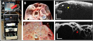
Optical coherence tomography (OCT) can distinguish between pancreatic cysts that are at low risk for becoming malignant and those that are high-risk. Other optical techniques often fail to provide images that are clear enough to differentiate between the two types. A team of researchers from four Boston-area institutions used OCT to examine pancreatic tissue samples from patients with cystic lesions. By identifying unique features of the high-risk cysts that appeared in the OCT scans, the group developed a set of visual criteria to differentiate between high- and low-risk cysts. They then tested the criteria by comparing OCT diagnoses to those obtained by examining thin slices of the pancreatic tissue under a microscope. Their results, described in the August issue of the journal Biomedical Optics Express, showed that OCT allowed differentiation between low- and high-risk cysts with a success rate close to that achieved by microscope-assisted examinations of slices of the same samples. Additional general information:
Additional technical information: |
A: Photograph of the OCT instrument and imaging probe; B,B’: Gross appearance and magnified OCT image of the malignant mucinous cystic neoplasm (MCN); C,C’: Gross appearance and magnified OCT image of the benign serous cystic adenoma (SCA). MCNs have large cavities filled with a mucinous fluid that scatters light (yellow arrow), while SCAs have a honeycomb structure of smaller cavities separated by tiny septae (red arrow). The SCA fluid is optically transparent and does not scatter light. OCT scale bar: 500 μm. (Images: Nicusor Iftimia, Physical Sciences Inc., and William Brugge, Massachusetts General Hospital) |
||
|
OCT is being used to differentiate between benign and |
|||


