|
|
 |
Larynx & Vocal Cord |
|
|---|---|
|
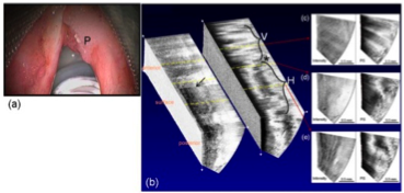
OCT has been used to image the larynx during surgical endoscopy. Office-based OCT has the potential to guide surgical biopsies, direct therapy, and monitor disease. This is a promising imaging modality to study the larynx. In-vivo 3D human vocal fold images have been demonstrated with polarization sensitive optical coherence tomography (PS-OCT), allowing characterization of the extent and location of vocal fold lesions, and thus providing useful information in guiding surgeons during phonomicrosurgery. Laser implications: The Insight laser's long coherence length and low polarization state wobble are particulary beneficial for PS-OCT applications. Additional technical information:
|
|
|
OCT allows the physician to see the internal structure |