|
|
 |
Fetal Heart Diagnostics |
|||
|---|---|---|---|
|
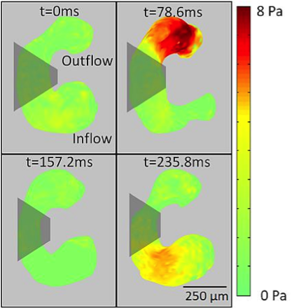
For the first time, researchers at Case Western Reserve University (Cleveland, Ohio), using an optical coherence tomography (OCT) method, have been able to visualize in 3D the stresses induced by flowing blood in an embryonic heart. The method is promising for learning how and why heart defects develop. In laboratory experiments, Rollins and colleagues directly measured the heart structure and blood flow within the developing hearts of quail embryos. learned from these preliminary animal tests and develop a way to apply this method to humans. The ultimate goal is to develop a tool that doctors can use to decide if early intervention could put a developing heart back on the right track, preventing a defect. Additional general information: Doppler OCT imaging sheds light on health of baby's heart Additional technical information: 4D shear stress maps of the developing heart using Doppler optical coherence tomography |
|
Stresses in an embryonic heart may predict future problems and determine whether or not early intervention is warranted. |
|