|
|
 |
Diagnosing Multiple Sclerosis |
|
|
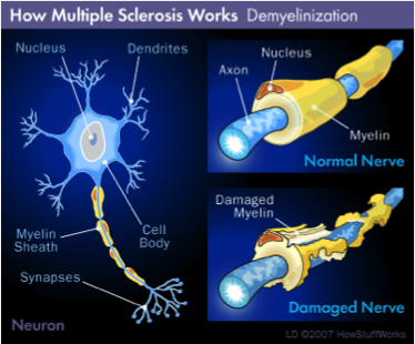
OCT is being investigated as a way to diagnose multiple sclerosis (MS). By imaging the very small nerves in the back of the eye, MS can be diagnosed earlier. As it leaves the eye, the optic nerve is the only tissue composed of unmyelinated axons. These can be imaged directly. The retinal nerve fiber layer (RNFL) is made up of the axons of the retinal ganglionar cells that convey the visual information from the retina to the lateral geniculate nucleus. Until they exit the eye, they do not acquire the protective myelin sheath. This extraordinary circumstance allows us to study the influence on isolated axons of several diseases. Ganglionar cells and their axons, besides being the main retinal component around the optic nerve (90% of retinal thickness) are also representative at the macula (30–35%). Both time-domain and spectral-domain optical coherence tomographies use light interference patterns to produce a tomogram, or cross-section, through the layers of the retina. The OCT software then constructs a two-dimensional (time-domain, TD-OCT) or three-dimensional (spectral-domain, SD-OCT) image of the retina and the optic nerve and is capable of measuring the different layers of the retina with a margin of error of 4–6 μm. The reproducibility of RNFL and macular measures is excellent with SD-OCT in multiple sclerosis. Multiple studies have provided information on the normal values for the RNFL thickness and have reported the RNFL loss that occurs after different pathologies that affect the optic nerve. Axonal loss, in contrast to demyelination, is not reversible and is therefore an important cause of sustained disability. In patients diagnosed with multiple sclerosis, it has been demonstrated that axonal loss occurs in the early stages of the disease. This is one of the reasons supporting the early use of neuroprotective drugs. Monitoring axonal loss has become a priority in multiple sclerosis and OCT as a sensitive, precise, and reproducible technique is acquiring increasing importance for both neurologists and ophthalmologists. Additional general information:
Additional technical information: |
Nerve deterioration can be detected using OCT.
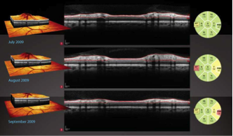
These OCT images track nerve deterioration over time. |
|
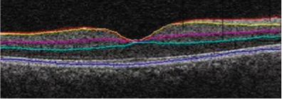
Using OCT to measure eye layer thicknesses |
|