|
|
 |
Dermatology |
|
|---|---|
|
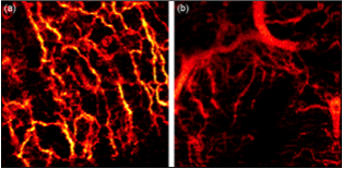
OCT is being used in dermatology to identify nonmelanoma skin cancer and diagnose conditions (such as psoriasis, eczema, and Bowen's disease) without taking a biopsy. An optical coherence tomography (OCT) technique that noninvasively maps the network of tiny blood vessels in the papillary dermis could soon help doctors better diagnose, monitor, and treat skin cancer. Scientists at Medical University Vienna (MUW) used a high-resolution three-dimensional imaging method to visualize the network of blood vessels beneath the outer layer of the skin–the epidermis–that feed cancerous lesions. “The condition of the vascular network carries important information on tissue health and its nutrition,” said Rainer Leitgeb, an MUW researcher and the study’s principal investigator. “Currently, the value of this information is not utilized to its full extent.” The network of vessels supplying blood to the tested lesions can show significantly altered patterns in comparison to healthy skin. Images of basal cell carcinoma have shown a dense network of unorganized blood vessels, with large vessels abnormally close to the skin surface. The larger vessels branch into secondary vessels that supply blood to energy-hungry tumor regions. OCT images, together with information about blood flow rates and tissue structure, could yield important insight into the metabolic demands of tumors during different growth stages. OCT can help assess how quickly tumors grow and spread, as well as monitor treatment effectiveness. Laser implications: Insight’s fast measurement speed reduces movement blurring and supports high definition 3D imaging. Long coherence length allows OCT system design flexibility to better map blood flow velocity. Our clean optical signal provides more detail, higher contrast and less feature blur. Additional general information: Additional technical information: |
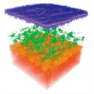
3D image of a fingertip, including sweat glands.
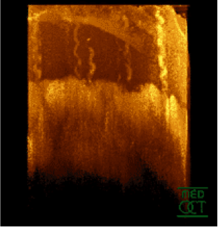
Segmented optical coherence tomography image of |
|
Healthy versus diseased skin vasculature: (a) A healthy network of blood vessels in the |
|