|
|
 |
OCT Technology |
|
|---|---|
|
Optical Coherence Tomography (OCT) is a technique for obtaining subsurface images of translucent or opaque materials at a resolution equivalent to a microscope. Effectively, it is "optical ultrasound" imaging reflections from within tissue to provide cross-sectional images. OCT is being applied across the medical community because it provides tissue morphology imagery at much higher resolution (better than 10 µm) than other imaging modalities such as MRI or ultrasound. The key benefits of OCT are:
OCT delivers high resolution because it is based on light rather than on sound or radio frequency. An optical beam is directed at the tissue, and a small portion of light that reflects from subsurface features is collected. Note that most light is not reflected but scatters off at large angles. In conventional imaging, this diffusely scattered light contributes to background that obscures an image. In OCT, however, optical coherence is used to record the optical path length of received photons, thus allowing rejection of most photons that scatter multiple times before detection. This is why OCT can build up clear 3D images of thick samples by rejecting background signal while collecting light directly reflected from surfaces of interest. Within the range of noninvasive 3D imaging techniques that have been introduced to the medical research community, OCT as an echo technique is similar to ultrasound imaging. Other medical imaging techniques such as computerized axial tomography, magnetic resonance imaging, or positron emission tomography do not utilize the echo-location principle. The technique is limited to imaging 1 to 2 mm below the surface in biological tissue because at greater depths the proportion of light that escapes without scattering is too small to be detected. No special preparation of a biological specimen is required, and images can be obtained ‘non-contact’ or through a transparent window or membrane. It is also important to note that the laser output from the instruments is low (eye-safe near-infrared light is used) and damage to the sample is, therefore, unlikely. The level of detail provided by its high spatial resolution makes OCT suitable for investigating structures that are not revealed by other imaging methods. For example, in cardiovascular research OCT offers the potential to image in great detail not only the surface of an artery but also the layers beneath the surface of the vessel. OCT with its spatial resolution in the micrometer range offers the possibility to investigate the structural details of an artery to detect the presence of atherosclerotic disease. NRC’s Institute for Biodiagnostics, the Industrial Materials Institute, and the Institute for Microstructural Sciences are developing OCT hardware systems to target medical applications in cardiac and oral health.
Additional general information:
|
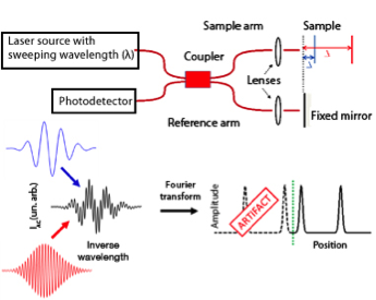
An OCT system diagram and response.
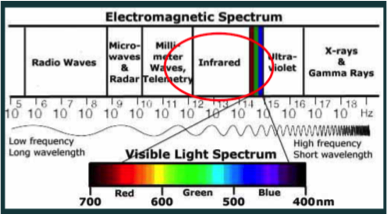
OCT uses light in the near-infrared region. |